
Our overarching goal is to understand the basic mechanisms that support learning and memory and how these mechanisms become hijacked in epilepsy. We have several exciting research projects that utilize cutting-edge techniques to gain new insights into brain function and pathology.

Hypothalamic control of hippocampal function
The supramammillary nucleus of the hypothalamus extends axons directly to the hippocampus and higher order cognitive centers, but its function has remained poorly understood. We recently found that the supramammillary exerts potent and cell type specific control of locomotion and broadcasts a “speed signal” to the hippocampus.
An additional ongoing project which is funded by NIH through the K99/R00 mechanism is to determine the role of the mammillary bodies, which connect to the hippocampus via Papez circuit, in controlling network synchrony in epilepsy. This area is composed of multiple cell types with distinct projections and can control synchronous network states (see below) - a role we hypothesize is pathologically hijacked in epilepsy to promote seizure activity.

Network mechanisms for memory
Neural activity is typically fragmented across various timescales, which is thought to support distinct brain states and modes of communication. The organizational principles that support the compression of neural activity into network rhythms and synchronous population events is poorly understood. We study the interplay between local microcircuits and large-scale brain networks as it relates to behaviorally relevant network dynamics. In particular, we are interested in two highly synchronous events which are named after their distinct waveform features: the sharp-wave ripple recorded in area CA1 and the dentate spike recorded in the dentate gyrus. Unlike the sharp-wave ripple, which supports memory consolidation through replay events, a role for the dentate spike is unknown. Our lab seeks to understand the basic functions of dentate spikes and their potential role for creating pathological synchrony in epilepsy. See Farrell et al., Nature (2024)

In vivo visualization of endocannabinoid signaling
The endocannabinoid CB1 receptor is the most abundant GPCR in the brain and potently shapes synaptic activity in an activity-dependent manner. The endogenously synthesized lipid ligands are extremely difficult to study in the brains of awake behaving mouse, leading to many unresolved questions about which endocannabinoid contributes to activity-dependent CB1 signalling? Where? For how long? We overcame this limitation with a genetically encoded endocannabinoid sensor and characterized this tool in vivo (see Dong et al., Nature Biotech 2022).
Using this sensor, we then showed that the endocannabinoid 2-AG is overproduced during seizures, broken down into arachidonic acid, thus providing substrate for COX-2 to generate pathologically high prostaglandin levels (see Farrell et al., Neuron 2021). This COX-2 pathway has profound effects on the neurovascular system, mimicking a stroke, and leads to prolonged behavioral impairment after seizures (see Farrell et al., eLife 2016). Thus, we are interested in how endocannabinoids not only shape neural activity, including during seizures, but how this metabolic crosstalk with prostaglandins regulates cerebral blood flow.

Non-invasive cell type-specific neuromodulation
Low intensity focused ultrasound (fUS) is an emerging approach to influence brain activity via non-invasive ultrasound waves. The translational potential is massive, but only if we understand how the endogenous circuit dynamics can be selectively tuned to counteract brain pathophysiology. We previously published a new tool that can monitor the activity of genetically-defined cell populations with fiber photometry to understand how various fUS parameters can be modified to recruit specific cell types and ultimately disrupt seizures (see Murphy et al., PNAS 2022). In our lab, we will push this technology further, working towards resolving sub-cellular dynamics in vivo across a range of cell types and brain areas with the goal of guiding future clinical interventions.
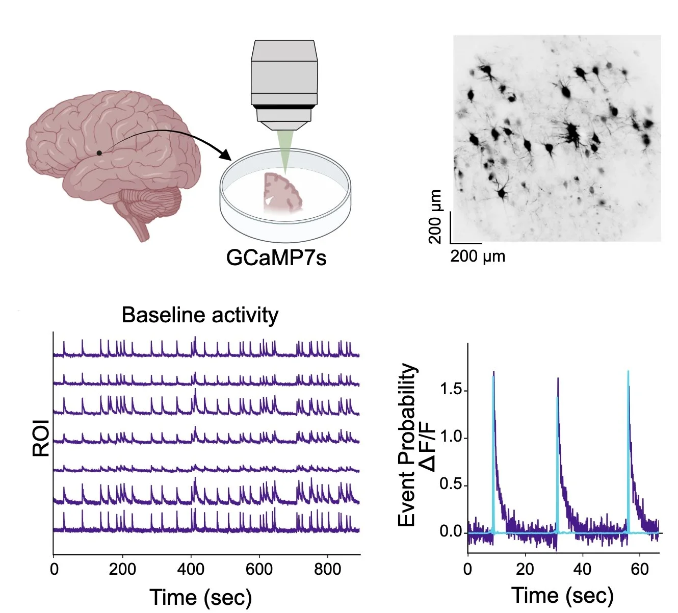
Interrogating human cortical circuits
In close collaboration with Dr. Emily Osterweil’s lab, we have leveraged resected cortical tissue from Boston Children’s to understand the organization and function of human circuits. Using slice culture approaches to keep tissue healthy for weeks, we are able to perform genetic perturbations and express genetically-encoded tools to manipulate gene expression and understand the molecular, cellular, and circuit-level consequences. We validated this approach for studying the functional role of FMR1, the gene underlying the most common genetic cause of autism (Singh et al., BioRxiv 2026), but this framework can be applied to many other genetically-driven disorders, which we are actively pursuing (e.g., SHANK3, SCN1A, ANKRD17, TSC2, SYNGAP1, and others). Moreover, we are constantly expanding our toolkit (borrowed from our in vivo mouse research) to interrogate human circuits - utilizing calcium imaging, optogenetics, voltage imaging, biosensor assays, and other approaches.
Funding support has generously been provided by: